Why does QRS complex have the largest amplitude?
 Michael Henderson
 Thereof, why is the QRS complex so much larger than the P wave?
The QRS complex indicates ventricular depolarization. Depolarization triggers contraction of the ventricles. Because of the larger tissue mass, the QRS complex is larger than the P wave.
Furthermore, what does a widened QRS complex indicate? A “wide QRS complex” refers to a QRS complex duration ≥120 ms. Widening of the QRS complex is related to slower spread of ventricular depolarization, either due to disease of the His-Purkinje network and/or reliance on slower, muscle-to-muscle spread of depolarization.
In this regard, why does the first heart sound occur after the QRS complex?
This depolarization of ventricular cells underlies the QRS complex within the ECG. As the ventricular cells contract, intraventricular pressures increase above those in the atria, and the atrioventricular valves abruptly close. Closure of the atrioventricular valves results in the first heart sound, S1.
Why might a very fit person have a slower heart rate than someone of average fitness?
Their resting heart rate is lower because a fit person has a larger stroke volume. his means that their heart pumps a larger volume of blood per second. They have a greatervolume of oxygen that is delivered to the body per heartbeat.
Related Question Answers
What is the normal duration of a QRS complex?
The normal duration (interval) of the QRS complex is between 0.08 and 0.10 seconds — that is, 80 and 100 milliseconds. When the duration is between 0.10 and 0.12 seconds, it is intermediate or slightly prolonged. A QRS duration of greater than 0.12 seconds is considered abnormal.Why is the QRS complex upside down?
The QRS complex is ventricular depolarization. It is typically much wider than the ventricular depolarization that generates the QRS. Sometimes it is upside down (inverted). Sometimes half of it is upside down and the other half upright; this is called biphasic.What position is best for electrodes?
Right sided ECG electrode placement- The most useful lead is V4R, which is obtained by placing the V4 electrode in the 5th right intercostal space in the mid-clavicular line.
- ST elevation in V4R has a sensitivity of 88%, specificity of 78% and diagnostic accuracy of 83% in the diagnosis of RV MI. [ see Inferior STEMI]
What do the P QRS and T waves represent?
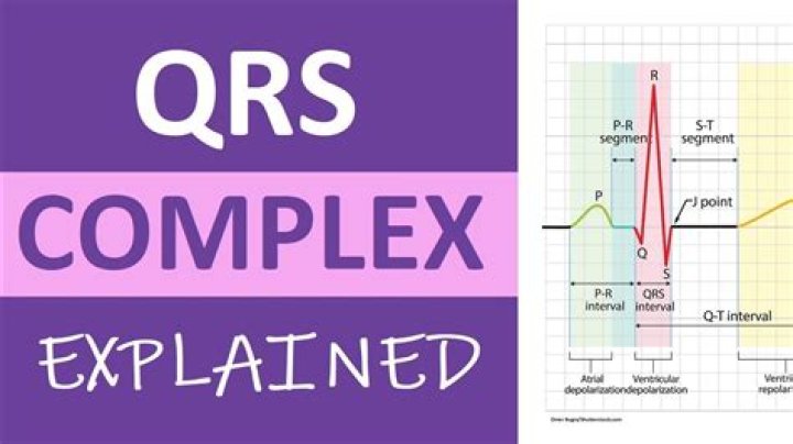
ECG – A Pictorial Primer. Atrial and ventricular depolarization and repolarization are represented on the ECG as a series of waves: the P wave followed by the QRS complex and the T wave. The first deflection is the P wave associated with right and left atrial depolarization. The second wave is the QRS complex.How do you read a QRS complex?
Here are some simple steps on how to measure the QRS complex:- Find the QRS on the EKG strip.
- Determine where the QRS complex is, and to do this you start measuring the END of the PR interval to the END of S-wave.
- Count the SMALL boxes between there measurements.
- Remember each box represents 0.04 seconds.
What happens during the QRS complex?
As the name suggests, the QRS complex includes the Q wave, R wave, and S wave. These three waves occur in rapid succession. The QRS complex represents the electrical impulse as it spreads through the ventricles and indicates ventricular depolarization.Why is there no atrial repolarization wave?
There is no distinctly visible wave representing atrial repolarization in the ECG because it occurs during ventricular depolarization. Because the wave of atrial repolarization is relatively small in amplitude (i.e., has low voltage), it is masked by the much larger ventricular-generated QRS complex.What can you say about the amplitude of various waves in different cardiac cycles?
What can you say about the amplitude of the various waves in different cardiac cycles? The P wave and the QRS complex represent depolarization of the atrial and ventricular muscle respectively. The heart muscle is also stronger, meaning it pumps more blood with each successive beat so it has to beat less often.Why is there a time delay between the QRS complex and peak blood flow?
Why is there a time delay between QRS complex and peak blood flow (volume pulse) in finger? The QRS is the electrical signal telling the ventricle to contract. It takes a little time for the heart to contract, and for the pressure wave to make its way to the finger.How does the amplitude of the P wave compare to that of the QRS complex?
The P wave describes the depolarization of the right and left atria. The amplitude of this wave is relatively small, because the atrial muscle mass is limited. It has a smaller amplitude, compared to the QRS complex, and is usually observed 300 ms after this larger complex.Is lub systolic or diastolic?
The “ lub” is the first heart sound, commonly termed S1, and is caused by turbulence caused by the closure of mitral and tricuspid valves at the start of systole. The second sound,” dub” or S2, is caused by the closure of aortic and pulmonic valves, marking the end of systole.Can an EKG show ventricular contraction?
Unlike contraction of the atria, ventricular contraction can be confirmed clinically by palpating a pulse or by monitoring a pulse oximeter wave form. A patient in cardiac arrest may have normal QRS complexes on his or her ECG; ventricular muscle cells are depolarizing, but there is no contraction.What would be the heart rate of a person?
Your pulse rate is the number of times your heart beats per minute. A normal resting heart rate should be 60–100 beats per minute, but it can vary from minute to minute.What is systole and diastole?
Your systolic blood pressure is the top number on your reading. It measures the force of blood against your artery walls while your ventricles — the lower two chambers of your heart — squeeze, pushing blood out to the rest of your body. Your diastolic blood pressure is the bottom number on your reading.Which heart sound is produced as the AV valves close?
Heart SoundsThe first heart sound (S1) represents closure of the atrioventricular (mitral and tricuspid) valves as the ventricular pressures exceed atrial pressures at the beginning of systole (point a).
What do you think causes heartbeat sounds?
The sound of a heartbeat is caused by the heart valves opening and closing as they pump blood. When the heart is working properly, blood can only flow in one direction. The valves make this possible by opening and closing in exact coordination with the heart's pumping action.How do you treat a wide QRS complex?
If QRS width greater than or equal to 0.12 seconds, assess if the rhythm is regular (and obtain expert consultation). For regular WCTs, if VT or uncertain rhythm, amiodarone; prepare for elective synchronized cardioversion. If SVT with aberrancy, treat with IV adenosine (vagal maneuvers).What electrolyte imbalance causes widened QRS complex?
ECG characteristics of hyperkalemia, high blood potassium:- P-waves are widened and of low amplitude due to slowing of conduction.
- QRS complex: QRS widening. fusion of QRS-T. loss of the ST segment.
- Tall tented T waves.
What is the treatment for wide complex tachycardia?
For a stable and regular wide-complex tachycardia, if ventricular, amiodarone 2 × 150 mg intravenously over 20–60 minutes is a safe treatment of choice. In previously known left ventricular fascicular ventricular tachycardia, verapamil and a beta-blocker are first-line options.How is the QRS complex described in atrial fibrillation?
Frequently, coarse “fibrillatory waves” can be seen representing the erratic atrial activity that occurs in the setting of atrial fibrillation. The QRS complexes are “irregularly irregular” as there is no pattern to their frequency. This is commonly described as varying R-R intervals.How many boxes is a wide QRS?
Wide refers to a QRS complex duration (width) of greater than or equal to 0.12 seconds (120 msec), corresponding to three small boxes on the ECG paper.What sport has the highest heart rate?
SwimmingWill my heart rate decrease as I get fitter?
Athletes often have a lower resting heart rate than others. If you exercise frequently and are reasonably fit, your heart rate may be lower than other people. This isn't necessarily a bad thing. A low heart rate means your heart needs fewer beats to deliver the same amount of blood throughout your body.What heart rate is a heart attack?
Can your heart rate reveal your risk for a heart attack? A very high or very low heart rate may reveal your risk for heart attack. For most people, a heart rate that's consistently above 100 beats per minute or below 60 beats per minute for nonathletes should prompt a visit to a doctor for a heart health evaluation.Is a resting pulse of 56 good?
The normal range is between 50 and 100 beats per minute. If your resting heart rate is above 100, it's called tachycardia; below 60, and it's called bradycardia. Increasingly, experts pin an ideal resting heart rate at between 50 to 70 beats per minute.What happens if your heart rate is too high?
When your heart is beating too fast, it may not pump enough blood to the rest of your body. This can starve your organs and tissues of oxygen and can cause the following tachycardia-related signs and symptoms: Shortness of breath. Lightheadedness.What causes resting heart rate to decrease?
Your heart beats faster to accelerate your blood circulation and so regulate your body temperature. Conversely, when you're in a cooler environment, the blood circulation in peripheral parts of the body decreases. Your heart has less work to do and your resting heart rate will decrease.Can you damage your heart by exercising too hard?
Pushing your body to the max day after day can stress your heart and raise your risk for a type of abnormal heart rhythm called atrial fibrillation, or A-fib, which ultimately can lead to heart failure or a stroke, according to the review, which analyzed 12 studies on A-fib in athletes and endurance runners.What is a good resting heart rate by age chart?
Children have higher resting heart rates than adults- Newborns 0 to 1 month old: 70 to 190 bpm.
- Infants 1 to 11 months old: 80 to 160 pm.
- Children 1 to 2 years old: 80 to 130 bpm.
- 3 to 4 years old: 80 to 120 bpm.
- 5 to 6 years old: 75 to 115 bpm.
- 7 to 9 years old: 70 to 100 bpm.
- 10 years and older: 60 to 100 bpm.